High-Frequency Ventilation
Objectives — Determine initial HFOV settings for neonatal and pediatric patients, and understand the principles, gas-transport mechanisms, and settings of high-frequency ventilation.
Principles of HFV
High-frequency ventilation is a form of invasive mechanical ventilation that uses small tidal volumes — less than anatomic dead space — at rapid frequencies, sometimes greater than 900 breaths/min.
Primary goal: provide adequate ventilation and oxygenation while limiting lung injury.
How HFV Oxygenates
Oxygenation is achieved by maintaining a high lung resting volume (increased FRC) through a high mean airway pressure (Paw), similar to CPAP, between 16–30. This is essentially a recruitment maneuver — maintaining higher alveolar pressure for better gas exchange and improving the V/Q ratio by opening previously collapsed alveoli.
HFV reduces overstretching of the alveoli at high pressures and the repetitive opening/closing stresses on the lungs, which reduces ventilator-induced lung damage. (Think "CPAP with a wiggle.")
Two Types
| Type | Notes |
|---|---|
| HFOV — High-Frequency Oscillatory Ventilation | The most common form of HFV |
| HFJV — High-Frequency Jet Ventilation | Used in pediatrics/neonates, mostly for BPD, PIE, and air leak syndromes |
Indications for HFV
- Failure of conventional ventilation (CMV)
- Preterm infants with severe lung disease requiring PIP > 30 cmH₂O
- Severe meconium aspiration syndrome
- Persistent pulmonary hypertension not responding to maximum support
- Congenital diaphragmatic hernia
- Air leak syndromes (pneumothorax, PIE)
- Severe RDS, pulmonary hypoplasia
Quantitative thresholds for failure of conventional ventilation: plateau pressures ≥ 30–35 cmH₂O with Vt of 5–7 mL/kg and severe respiratory acidosis (pH < 7.1). For failure of oxygenation (e.g., ARDS): SpO₂ < 90%, PaO₂/FiO₂ < 150 despite FiO₂ > 60% and optimal PEEP, or an oxygenation index (OI) > 15.
OI = (MAP × FiO₂%) / PaO₂
Gas Transport Theory of HFV
How does air exchange happen when the tidal volume is smaller than the dead space? Five mechanisms work together.
| Mechanism | What it does |
|---|---|
| Spike formation | A high-energy waveform penetrates the center of the airway lumen, enhancing bulk flow of fresh gas deeper into the lungs |
| Helical diffusion | Gas exiting along the circumference of the airway (passing the spike) carries CO₂ out as fresh gas enters through the middle |
| Taylor dispersion | The augmented diffusion of gas in parabolic flow — fresh gas in through the center, CO₂ out along the walls, balancing diffusion |
| Pendelluft ventilation | Gas mixing between lung regions with different time constants — fast units fill quickly and, at end-inspiration, empty into the slow-filling units |
| Molecular diffusion | The rapid random thermal oscillation of molecules; occurs as long as temperature is constant, allowing gas exchange at the A/C membrane |
HFOV Settings
HFOV uses a piston of microprocessor gas controllers to push air into and out of the lungs. Fresh gas is delivered through the circuit as bias flow. Settings are patient-dependent based on size, weight, and disease process.
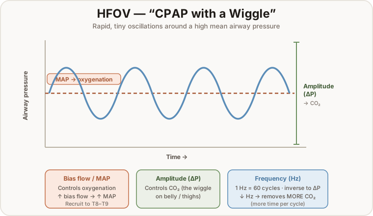
Bias Flow
- Flow at a constant rate.
- Helps sweep out excess CO₂ and provides the flow to generate MAP.
- Increasing bias flow → increase in MAP (impacts PaO₂ upward).
- Adjusts both oxygenation and CO₂ elimination.
MAP (Mean Airway Pressure)
- Provides oxygenation and lung-recruitment pressure.
- Increase until lung expansion reaches the thoracic vertebrae T8–T9; maintain MAP once inflated to T8–T9.
- Can be set from conventional ventilation by adding +5 to the MAP currently required to ventilate/oxygenate.
Amplitude (∆P)
- The tidal volume — controls CO₂ in the same manner.
- The oscillatory pressure amplitude creates the "wiggle" on the patient's belly, thighs, or xiphoid process.
Frequency (Hz)
- One Hz = 60 breath cycles.
- Frequency has an inverse effect on amplitude — as frequency increases, amplitude decreases (smaller time to deliver the tidal volume).
- To eliminate more CO₂, decrease the frequency — allowing more time for pressure to reach the alveoli.
I-time & FiO₂
- I-time: generally set to 33% and not adjusted.
- FiO₂: set by a blender attached to the vent; follow similar O₂ settings.
HFJV Settings
High-frequency jets operate on the principle of a nozzle or injector that creates high-velocity "jets" of gas directed into the lungs. Exhalation is passive, using the elastic recoil of the lungs. HFJV is used only in pediatrics and neonates, mostly for BPD, PIE, and other air leak syndromes.
| Setting | Value / role |
|---|---|
| I-time | Generally 0.02 sec — minimal time for the jet to cause large pressure changes in the alveoli (protects against VILI) |
| Rate | 240–660 to meet the patient's requirements |
| PIP | Set to maintain a driving pressure (∆P = PIP − PEEP) — important for alveolar ventilation and CO₂ elimination; can be set lower than the PIP on the vent |
Oxygenation vs. Ventilation on HFJV
Conventional ventilation drives oxygenation when paired with the jet:
- Increase PEEP to improve oxygenation (often higher PEEP than normal CMV).
- Increase the CV rate (a lung-recruitment maneuver — generally means PEEP is too low).
- Increase I-time.
HFV settings are primarily for ventilation — delta P is the most important factor for changing PaCO₂:
- Increase PIP on the HFV to decrease PaCO₂.
- Decrease PEEP.
- Adjust rate between 240–660 BPM.
Servo Pressure
Servo pressure is the driving pressure that automatically regulates flow. It changes as lung volume or mechanics change, acting as an early warning that the patient is getting better or worse.
| Servo pressure increases with | Servo pressure decreases with |
|---|---|
| Improving compliance or resistance | Worsening compliance and resistance |
| Leak around the ETT (baby is growing) | Obstructed ETT |
| Tubing leak | Tension pneumothorax |
| Right mainstem intubation |