The Newborn Assessment & RDS
Objectives — Identify RDS in the infant through maternal and infant assessments: evaluate the newborn at delivery, apply the correct resuscitation and oxygen targets, and recognize the signs, pathophysiology, and treatment of respiratory distress syndrome.
Evaluation of the Newborn
All newborns should be assessed immediately upon delivery. The team in the OR often consists of a doctor, a nurse, and an RT for immediate resuscitation of the newborn.
Two categories of infants will most likely need interventions (more than 90% will not):
- Infants with evidence of meconium
- Premature infants
Babies should be dried and warmed, placed into an incubator, and immediately assessed.
Lung Development
| Weeks of gestation | Event |
|---|---|
| 17–26 | Airways increase in length and diameter |
| 24–25 | Vascularization of an adequate capillary system around the lungs occurs |
| ~23–26 | The distance between primitive alveoli and capillaries is close enough for gas exchange — infants become viable and can be supported |
Meconium Aspiration (Overview)
Meconium is the infant's first stool/bowel movement — a very sticky green-black substance that, if inhaled, can cause significant respiratory problems. (The full disease process is covered in the disease-processes lesson.)
Resuscitation of the Newborn
In high-risk births, the RT is often part of the team caring for the infant right after birth. The most important action is effective ventilation, delivered with effective bag-mask ventilation.
If a premature infant has a heart rate less than 100 beats/min or is apneic, apply bag-mask ventilation first, at a rate of 40–60/min, with room air.
During emergency resuscitation of a premature infant, use the lowest oxygen concentration possible:
| Gestational age | Starting FiO₂ |
|---|---|
| > 35 weeks | 21% |
| < 35 weeks | 30% |
Effective positive pressure ventilation (PPV) usually results in rapid improvement of heart rate.
- Initial pressures for bag-mask resuscitation may be 30–40 cmH₂O, particularly for premature deliveries.
- If the heart rate continues to drop below 60/min, apply PPV first for 30 seconds.
- If there is no recovery within the first 30 seconds, begin chest compressions.
Scoring & Classification
APGAR Score
An objective scoring system used to evaluate a newborn rapidly, assigned at 1 minute and 5 minutes of life.
| Score | Interpretation |
|---|---|
| 7 or higher | Normal |
| ~7 | May need some intervention |
| < 6 after 1 minute | May require more aggressive support |
Ballard Maturational Assessment (BMA)
Used to determine gestational age by using six physical and neurological signs to assign a week of gestational age to the newborn.
Weight Classifications
| Classification | Weight |
|---|---|
| Low birth weight (LBW) | < 2500 g |
| Very low birth weight (VLBW) | < 1500 g |
| Extremely low birth weight (ELBW) | < 1000 g |
Weights can be compared to the Ballard scoring system to define the developmental status of the child.
Colorado Intrauterine Growth Chart
Based on a large population of infants, comparing weights to gestational age to produce a growth curve. It estimates whether a baby is developing properly, or gives a quick estimate of gestational age from weight.
| Percentile | Classification |
|---|---|
| 90th and above | LGA — large for gestational age |
| 10th and below | SGA — small for gestational age |
Small preterm infants are at the highest risk: lungs unprepared for gas exchange (RDS), poor fat absorption, weak immune systems, a large surface-area-to-weight ratio (heat loss, impaired thermoregulation), and underdeveloped vasculature (increased hemorrhage risk, especially in the developing ventricles of the brain).
Vital Signs
Respiratory Rate
- Normal infant RR is 40–60 breaths/min.
- The lower the gestational age, the higher the likely respiratory rate.
- A rate > 60 is considered tachypnea.
- Breaths < 40/min should be checked against the child's baseline.
Heart Rate & Pulses
- Normal infant HR is 100–160 beats/min.
- Assess by auscultation of the apical pulse, normally at the fifth intercostal space, midclavicular line.
- Check the femoral and brachial pulse sites. Weak pulses indicate hypotension, shock, or vasoconstriction.
| Pulse finding | Significance |
|---|---|
| Bounding peripheral pulses | Major left-to-right shunting through a patent ductus arteriosus (PDA) |
| Strong brachial, weak femoral | May suggest PDA or coarctation of the aorta |
Silverman Scoring System
An identifier for the severity of lung disease after birth. Auscultation of the infant chest is difficult due to size and sound transfer, so breathing difficulty is judged by observing key physical signs:
- Nasal flaring
- Cyanosis
- Expiratory grunting
- Tachypnea
- Retractions
- Paradoxical breathing
Respiratory Distress Syndrome (RDS)
RDS is expressed immediately after birth. It is most likely due to insufficient surfactant.
Clinical Signs
- Tachypnea with worsening retractions, paradoxical breathing, audible grunting, and nasal flaring
- Increased fluid and atelectasis → decreased breath sounds or fine crackles on auscultation
- Cyanosis may or may not be noted
- Chest x-ray is definitive: diffuse, hazy, reticulogranular ("ground glass") densities with air bronchograms and low lung volumes
What the Signs Mean
| Sign | Mechanism |
|---|---|
| Nasal flaring | Decreases airway resistance and reduces airway collapse during the negative force of inhalation |
| Grunting | Splints the airway by forcing air against a partially closed glottis — an indicator of alveolar collapse |
| Retractions (suprasternal, substernal, intercostal) | Increased work of breathing, especially with decreased pulmonary compliance |
| Paradoxical breathing | The chest wall draws in while the abdomen stays put — a see-saw pattern indicating increased WOB |
Cyanosis can be absent despite severe hypoxemia. With high fetal hemoglobin and hyperbilirubinemia, an infant may appear normal in color even at a PaO₂ of 30 mmHg.
Pathophysiology (the Snowball Effect)
Major factors causing RDS: surfactant deficiency (below 34 weeks), decreased alveolar surface area (lung collapse, decreased compliance), increased small-airway compliance, and the presence of the ductus arteriosus.
To compound the primary problem of low surfactant, low oxygenation and ventilation increase pulmonary vascular resistance. Rising PVR leads to further right-to-left shunting that worsens as hypoxemia worsens (PPHN). Both hypoxemia and the developing acidosis from ineffective ventilation cause further surfactant reduction and worsening lung compliance.
Treatment
- CPAP and PEEP are both adequate means to improve outcomes in RDS.
- Advanced modes (HFV) may be required with rapid deterioration.
- Optimizing PEEP/CPAP improves oxygenation and FRC and ventilation.
Surfactant: Deficiency & Therapy
Surfactant is produced at 24 weeks of gestation. Prior to 34–35 weeks, the amount is adequate but trapped inside the alveolar type II cells, so it cannot be released to lower the surface tension of the alveoli (think LaPlace's law — high surface tension, decreased compliance).
The cascade:
- Surfactant deficiency → decreased lung compliance and high surface tension → alveolar collapse, atelectasis, increased WOB.
- The increased surface tension pulls fluid from the pulmonary capillaries, further decreasing compliance.
Surfactant Replacement
There are three surfactant preparations in the United States, generally derived from bovine or porcine sources. Some centers use prophylactic surfactant for infants under 1500 g. All surfactants are delivered via the ETT for rapid distribution. Infants are positioned on their sides per the manufacturer's guidelines, but may receive it supine if they cannot be repositioned.
| Drug | Generic / source | Dose (mg/kg phospholipid) | mL/kg | Dosing interval |
|---|---|---|---|---|
| Survanta | Beractant | 100 | 4 | Every 6 hr or more often |
| Infasurf | Calfactant | 100 | 3 | Every 12 hr or more often |
| Curosurf | Poractant alfa | 100–200 | 1.25–2.5 | Every 12 hr or more often |
Do not memorize the chart values — know the three drug/brand pairs and that all are given via the ETT.
Blood Gas & Saturation Targets
Capillary blood gas sampling and pulse oximetry assess the newborn for both congenital heart defects and respiratory disorders.
| Population | SpO₂ target | PaO₂ target |
|---|---|---|
| Pre-term infants | > 88% | 40–70 mmHg |
| Term infants & pediatrics | > 95% | — |
Pre- and Post-Ductal Monitoring
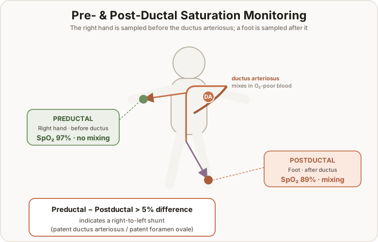
A patent ductus arteriosus and patent foramen ovale can cause right-to-left shunting, decreasing O₂ saturation from mixing. Detect it by placing one probe on the right arm (preductal) and one on the left lower leg (postductal):
A > 5% saturation difference between preductal and postductal is indicative of a right-to-left shunt — most probably a patent ductus arteriosus and patent foramen ovale.
Capillary Stick
Blood from a capillary stick on the heel of the foot is important for monitoring PCO₂ and electrolytes. Oxygen values from capillary sticks are inaccurate because it is a mixed venous sample.
If an infant has chronically high PCO₂ with normal oxygenation, a capillary stick is the best method of assessing PCO₂ status.
Pediatric Assessment
Assessment of pediatric patients is difficult due to age differences and rapid growth. Initial assessment begins with adequate perfusion and heart rate, starting with the patency of the airway.
- Heart rate and RR should be assessed against an age-appropriate chart.
- A respiratory rate of 20–40/min is generally acceptable for pediatric patients.
- A heart rate of 60–100 is acceptable (younger infants and toddlers may run higher).
- Asthma, allergic reactions, and infections are common — effective medication use is essential.
Recognizing Distress
- Accessory muscle use, grunting, flaring, and retraction are signs of respiratory distress (broadly called RDS).
- Head bobbing, chin up, and abdominal expansion are signs of impending respiratory failure.
- Levels of alertness — fully awake, agitated, minimally responsive, unresponsive — indicate whether a child needs an advanced airway.
Newborns with certain cardiac anomalies are dependent on their intracardiac shunts (ductus arteriosus, ductus venosus, foramen ovale) to survive. Increased saturation promotes constriction of the ductus arteriosus — normally a good thing, but it may cause premature closure in infants with ductal-dependent congenital heart defects. For example, an infant with hypoplastic left heart syndrome relies on the patency of the ductus arteriosus for systemic blood supply.