Neonatal & Pediatric Foundations
Objectives — Identify basic facts and terms about neonatal and pediatric respiratory care, and begin identifying RDS in the infant through maternal and infant assessments.
Basic Terms
The word neonatal comes from neo ("new") and natal ("pertaining to birth or origin"). Neonatal care includes both mature and premature infants.
| Term | Definition |
|---|---|
| Gestation | The period between conception and birth during which the embryo or fetus develops in the uterus |
| Mature infant | Born between 38–42 weeks of gestation |
| Premature infant | Born before 38 weeks gestation — considered high risk |
| Congenital | A disease or physical abnormality that is present at birth (e.g., gastroschisis, omphalocele, patent ductus arteriosus) |
Neonatal respiratory care is difficult because of the anatomical differences of the newborn:
- Heart shunting
- Underdeveloped lungs in infants born prior to 38 weeks
- Weight and size differences
Pediatrics
Pediatric respiratory care is much like adult respiratory care, with established disease processes and ventilator management. The primary difference is where the child or adolescent is in their growth, which is constantly changing.
- Pediatrics can include patients anywhere from infant to 21 years of age.
- Some pediatric patients are followed by the pediatric team past the age of 21 if they had an established disease process during their pediatric period (e.g., cancer).
Maternal factors have a major effect on the health of the child at birth. Assessment of the mother should be done initially.
Fetal Circulation & the Three Shunts
There are several differences in heart anatomy and fetal circulation. Three important bypass pathways function in the developing fetus to enhance the flow of blood to developing organs:
| Shunt | Location | Purpose |
|---|---|---|
| Ductus venosus | Between the liver and the inferior vena cava | Routes oxygenated blood from mom quickly back to the heart |
| Ductus arteriosus | Between the pulmonary artery and the aorta | Shunts blood away from the non-functional lungs |
| Foramen ovale | Between the two atria (through the interatrial septal wall) | Shunts blood right → left to reach the systemic circulation |
If these anatomical shunts remain open after birth, they cause a mixing of oxygenated and deoxygenated blood, which causes problems in the infant.
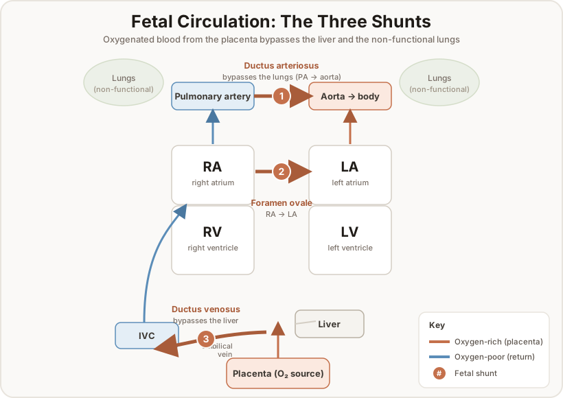
Ductus Venosus
A shunt between the liver and the inferior vena cava. It takes two-thirds of the oxygenated blood from the liver and allows it to make it back to the heart quickly to reach the rest of the body. This shunt closes rapidly after placental blood flow ceases — clamping the umbilical cord after birth helps close it.
Ductus Arteriosus
The ductus arteriosus allows shunting of blood away from the non-functional lungs in utero. It is located between the pulmonary artery and the aorta. In utero, pulmonary vascular pressure is higher than systemic vascular pressure, allowing most of the blood to shunt away from the lungs to the rest of the body.
This is normal and necessary — the lungs have no ability to oxygenate or ventilate, so the infant needs the oxygenated blood coming from the placenta to meet its systemic oxygenation needs.
This shunt normally closes after birth: as PaO₂ and PaCO₂ change in the pulmonary capillaries, the capillaries dilate, decreasing PVR and causing constriction of the ductus arteriosus.
The hormone prostaglandin can be given to maintain the patency of the ductus arteriosus after birth. This is useful in some disease processes among preterm infants (such as ductal-dependent congenital heart defects).
Prostaglandin
Prostaglandin is a hormone that slowly dissipates after the infant is born. Normally, prostaglandin levels keep the ductus open. As the smooth muscle of the blood vessels "senses" the drop in prostaglandin and increase in PaO₂, vessel constriction is triggered.
Foramen Ovale
A common shunt between the two atria that allows blood to shunt from the right to the left atrium through the interatrial septal wall. It allows oxygenated blood coming from mom (via the ductus venosus) to be shunted to the left side of the heart to quickly reach the systemic side of the infant's body.
This duct usually closes about three weeks after birth as post-utero circulation normalizes. Left-sided heart pressure (systemic vascular resistance, SVR) increases and helps close a flap of tissue over the foramen ovale, allowing it to seal.
Maternal Risk Factors
Maternal factors that could indicate adverse status of the neonate fall into three categories: medical, physical, and social.
| Maternal condition | Possible adverse outcomes |
|---|---|
| Previous pregnancy complications | Recurrence of complications |
| (Gestational) Diabetes Mellitus | Large gestational age (LGA), congenital malformations, RDS, hypoglycemia |
| Maternal age < 17 years | Low birth weight, prematurity |
| Maternal age > 35 years | Prematurity, chromosomal defects (Down syndrome) |
| Pregnancy-induced hypertension | Prematurity, SGA (small for gestational age), preeclampsia |
| Placenta previa | Placenta attaches over the cervix → prematurity, bleeding, SGA |
| Abruptio placentae | Placenta detaches from the wall / blood builds up behind it → fetal asphyxia, bleeding |
| Alcohol consumption | SGA, CNS dysfunction, mental retardation, facial dysmorphology |
| Smoking | SGA, prematurity, mental retardation, SIDS |
| Drug use | Placental abruption, IUGR, prematurity, CNS abnormalities, withdrawal disorders |
The Placenta
During the ~40 weeks of gestation, the placenta acts as the organ of respiration for the fetus — it extracts O₂ and excretes CO₂. Fetal hemoglobin and maternal hemoglobin exchange CO₂ and O₂. Placental attachment and placement can affect maternal factors for fetal development.
- Approximately 55 cm long
- 2 arteries, 1 vein
Fetal Assessment & Tests
Fetal assessment includes fetal scoring systems, ultrasonography, amniocentesis, fetal heart rate, and fetal blood gas analysis.
Amniocentesis
Direct sampling and quantitative assessment of amniotic fluid. Amniotic fluid can be used to detect meconium (fetal bowel contents), blood, and sloughed cells. Sloughed cells can be used to detect certain genetic abnormalities in utero.
Testing for Surfactant Deficiency
| Test | What it measures | Mature result |
|---|---|---|
| L/S ratio (Lecithin/Sphingomyelin) | Two phospholipids synthesized by the fetus; ratio increases with gestational age | 2:1 indicates stable surfactant production |
| Phosphatidylglycerol | A lipid found in pulmonary surfactant | > 1% = less than 1% chance of respiratory distress |
The L/S ratio uses amniotic fluid sampling to determine if the infant will have stable surfactant production at birth.
Fetal Heart Rate
Measurement of the fetal heart rate and uterine contractions during labor. Examining fetal heart rate changes related to uterine contractions identifies a fetus in distress.
- Normal fetal HR = 100–160 beats/min
- Tachycardia can indicate fetal hypoxemia, prematurity, or maternal fever
Decelerations are temporary declines in fetal heart rate:
| Severity | Decline |
|---|---|
| Mild | < 15 beats/min |
| Moderate | > 15 to 45 beats/min |
| Severe | > 45 beats/min |
There are early, late, and variable decelerations. Some are dangerous and could indicate a constriction on the baby's umbilical cord lifeline.
Umbilical Cord
The umbilical cord is the lifeline between mother and fetus. The thick gelatinous material that surrounds the umbilical arteries and vein is called Wharton's jelly. During fetal movement, the cord can pinch and interrupt blood flow — especially after the amniotic fluid is displaced during birth — causing decelerations and stressing the infant.
| Complication | Description |
|---|---|
| Cord prolapse | The cord passes through the cervix into the birth canal ahead of the presenting child. Compression inside the uterus is called an occult prolapse. |
| Nuchal cord | The cord wraps around the infant's neck — a dangerous scenario where the cord can compress the neck or be compressed upon delivery. |
Normal Birth / Delivery
Delivery is done one of two ways:
| Method | Notes |
|---|---|
| Vaginal delivery | Allows the baby to clear secretions by being "squeezed" through the birth canal |
| C-section | Cut through the abdominal wall to remove the baby; becoming a much more common delivery method |
Because a C-section skips the "squeeze," it is a setup for retained lung fluid — the basis of transient tachypnea of the newborn, covered in a later lesson.